Embarking on a comprehensive exploration of pal cadaver axial skeleton skull lab practical question 4, this discourse unveils the intricate anatomy of the skull, its significance within the axial skeleton, and its profound clinical applications. Prepare to delve into a captivating journey of anatomical discovery.
The skull, a remarkable structure composed of numerous bones, serves as the protective encasement for the brain and houses various sensory organs. Understanding its intricate composition is paramount in comprehending the human body’s intricate workings.
Anatomical Structures of the Skull: Pal Cadaver Axial Skeleton Skull Lab Practical Question 4

The skull, a complex and vital structure, serves as the protective encasement for the brain and houses various sensory organs. It comprises numerous bones, each playing a specific role in maintaining structural integrity and facilitating physiological functions.
Bones of the Skull
The skull consists of 22 bones, classified into two categories:
- Cranial Bones (8):Form the cranium, the protective vault enclosing the brain. These include the frontal bone, parietal bones, occipital bone, sphenoid bone, ethmoid bone, and temporal bones.
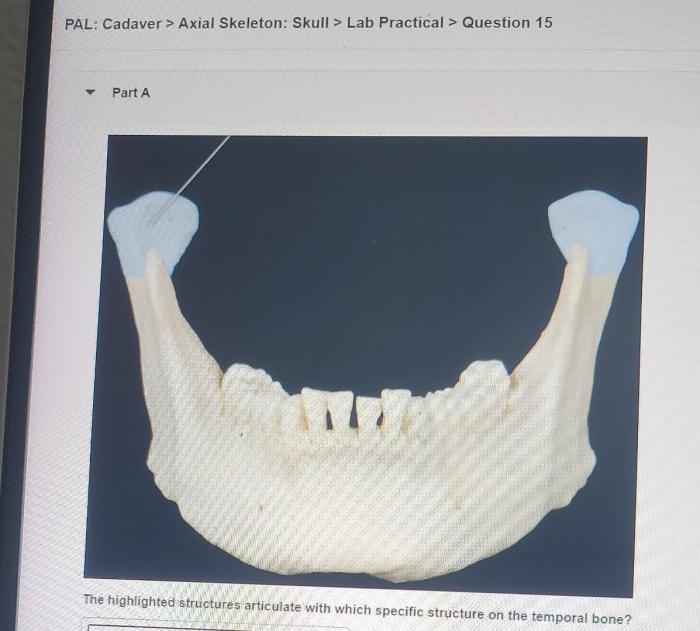
- Facial Bones (14):Form the facial skeleton, providing support for the facial features and housing the sensory organs. These include the nasal bones, maxillae, zygomatic bones, lacrimal bones, palatine bones, inferior nasal conchae, vomer, and mandible.
Sutures and Foramina
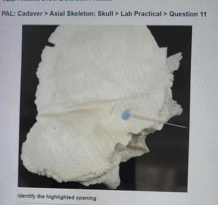
The bones of the skull are connected by fibrous joints called sutures, which allow for slight movement during growth and development. Foramina, openings in the skull, provide passageways for nerves, blood vessels, and other structures. Notable sutures include the coronal suture, sagittal suture, and lambdoid suture.
Important foramina include the foramen magnum, foramen ovale, and foramen rotundum.
Axial Skeleton of the Cadaver

The axial skeleton, a central component of the human skeletal system, provides support, protection, and mobility for the head, neck, and trunk. It comprises three main regions:
Components of the Axial Skeleton
- Skull:Encloses and protects the brain, housing the sensory organs.
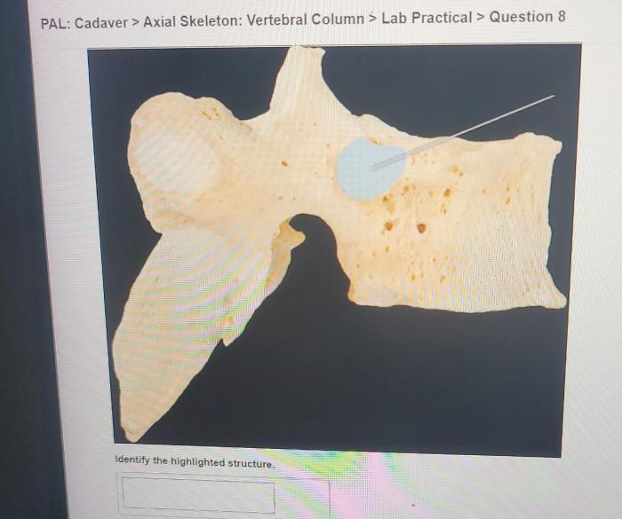
- Vertebral Column:A series of vertebrae stacked upon one another, providing support for the body, protecting the spinal cord, and allowing for movement.
- Rib Cage:Formed by the ribs and sternum, it protects the vital organs in the chest cavity, including the heart and lungs, and aids in respiration.
Functions of the Axial Skeleton
The axial skeleton serves crucial functions in the body:
- Support:Provides a rigid framework for the body, supporting the weight of the head and upper body.
- Protection:Encloses and safeguards delicate structures such as the brain, spinal cord, and vital organs.
- Mobility:Allows for movement of the head, neck, and trunk through the joints between the vertebrae and the skull.
Relationship between Skull and Vertebral Column, Pal cadaver axial skeleton skull lab practical question 4
The skull articulates with the first cervical vertebra, the atlas, via the atlanto-occipital joint. This joint allows for a wide range of head movements, including nodding, shaking, and tilting. The vertebral column extends inferiorly from the skull, providing support and protection for the spinal cord and allowing for flexibility and mobility of the neck and back.
Palpation of Skull Landmarks

Palpation, the process of feeling anatomical structures through the skin, is an essential skill in physical examination. Key anatomical landmarks on the skull can be palpated to assess its structure and identify potential abnormalities.
Palpable Landmarks
The following skull landmarks can be palpated:
- Frontal Bone:Smooth, convex surface at the forehead.
- Parietal Bone:Lateral to the frontal bone, forming the sides of the cranium.
- Temporal Bone:Located behind the parietal bone, housing the ear and containing the mastoid process.
- Occipital Bone:Forms the posterior part of the cranium, with the external occipital protuberance palpable at the back of the head.
Clinical Applications of Skull Anatomy
Understanding skull anatomy is essential in various healthcare fields:
- Neurosurgery:Surgeons rely on skull anatomy to plan and perform complex procedures on the brain and surrounding structures.
- Forensic Anthropology:Skull examination plays a crucial role in identifying human remains and determining age, sex, and ethnicity.
- Dentistry:Knowledge of skull anatomy is vital for diagnosing and treating dental problems related to the jaw and facial bones.
FAQs
What is the significance of understanding skull anatomy?
Understanding skull anatomy is crucial for various medical disciplines, including neurosurgery, forensic anthropology, and dentistry. It enables accurate diagnosis and treatment of medical conditions, aids in reconstructing facial features for identification purposes, and facilitates the design of dental prosthetics.
How does the skull contribute to the axial skeleton?
The skull forms the superior portion of the axial skeleton, providing protection for the brain and supporting the facial structures. It articulates with the vertebral column, allowing for head movements and providing stability to the body.
What are some key anatomical landmarks on the skull?
Palpable landmarks on the skull include the frontal bone, parietal bone, temporal bone, and occipital bone. These landmarks serve as reference points for surgical procedures, physical examinations, and anthropological studies.